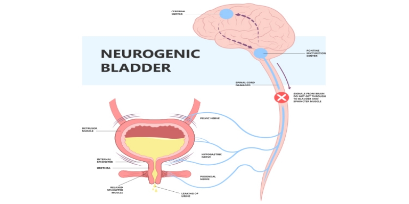
Best Female Neurogenic Bladder Treatment in India – RG Hospital Book Online Consultation International Patient Enquiry Quick Enquiry
X
Quick Enquiry
X
Choose File (File Format: Jpeg, Jpg, Png, PDF, Word Max. File Size: 2MB )
No file chosen...
All the fields are required
International Patient Enquiry
X
Choose File (File Format: Jpeg, Jpg, Png, PDF, Word Max. File Size: 2MB )
No file chosen...
All the fields are required
X
RG Marathon Championship 2022

Neurogenic Bladder

Neurogenic Bladder – Causes, Signs & Symptoms, Treatment, Management

The bladder is a hollow organ located in the pelvis, or lower abdomen. The bladder has two important functions:

  • It helps to store urine until it is full.
  • When the bladder becomes full, there is an urge to urinate, and the bladder empties completely without any leakage.

Urination can be abnormal if the bladder is unable to empty itself fully or empties itself before one reaches the bathroom. There can also be leakage before or after urination due to problems with bladder control.

When the conditions of the nervous system affect the bladder, it is called “neurogenic bladder.” Conditions like stroke, Parkinson’s disease, multiple sclerosis, disk herniation, and other nervous system abnormalities can result in neurogenic bladder.

There are two major types of bladder control problems that are associated with a neurogenic bladder. Depending on the nerves involved and nature of the damage, the bladder becomes either overactive (spastic or hyper-reflexive) orunderactive (flaccid or hypotonic).

  • People with overactive bladders have little to no control urinating. If the bladder is overactive, a person might feel a sudden urge to urinate or might go to the bathroom more often than necessary. An overactive bladder may result in incontinence (the involuntary or accidental release of urine).
  • Neurogenic underactive bladders behave in the opposite way. The bladder loses its ability to empty properly and fills beyond the normal capacity. At a certain point, the pressure of urine in the bladder overcomes the sphincter muscle’s ability to retain it, and urine leaks out. Underactive bladders fail to empty completely and hold on to urine.

What causes neurogenic bladder?

Several disorders can cause neurogenic bladder, including the following:

  • stroke
  • Parkinson’s disease
  • multiple sclerosis
  • spinal cord injuries
  • spinal surgeries
  • erectile dysfunction
  • trauma/accidents
  • diseases that affect the nervous system
  • central nervous system tumors
  • spinal congenital (present at birth) abnormalities
  • heavy metal poisoning

Underactive bladder may be a complication of diseases such as syphilis, diabetes, and polio.


What are the symptoms of neurogenic bladder?

The most common symptom of neurogenic bladder is being unable to control urination. Other symptoms include the following:

  • a weak or dribbling urinary stream
  • an inability to urinate or straining to urinate
  • frequent urination (urinating eight or more times daily)
  • urgency (a feeling or need to urinate immediately)
  • painful urination, which may mean there is a urinary tract infection

How is neurogenic bladder diagnosed?

Your doctor may order several tests of the nervous system and the bladder to diagnose neurogenic bladder:

  • Urodynamic studies (bladder function tests) are conducted to measure bladder capacity, bladder pressures, the flow of urine, and bladder emptying. A cystoscopy may be performed to examine the inside of the bladder and urethra (the tube through which urine passes) with a small telescope (cystoscope).
  • The skull, spine, and urinary tract may be examined with X-rays, computed tomography (CT), and magnetic resonance imaging (MRI). You may be referred to a neurologist for consultation.

The doctor will perform a physical examination and ask about your medical history, especially about any neurological problems such as back injury, stroke, or other neurologic diseases.


How is neurogenic bladder treated?

There are five types of treatment for neurogenic bladder: physical-psychological, electrical-stimulatory, drug therapy, intermittent self-catheterization, and surgery. The correct treatment is determined by symptoms, type, and extent of nerve damage, and a thorough discussion with the patient.


  • Physical-psychological therapy

    Physical-psychological therapy, also called timed voiding, combines will power and exercise. Your doctor will ask you to keep a voiding diary--a record of the amount and time you drink fluids, how many times you urinate each day, and whether you ever leak urine. This record creates a pattern that may help you determine the times of the day you should be near a bathroom, and the times when you should attempt to urinate. The periods of time between urination gradually get longer and longer as you gain control over urinating.

  • Electrical-stimulatory therapy In electrical-stimulatory therapy, electrodes and a small stimulator are placed near certain nerves during a minor surgical procedure. The stimulator is placed beneath the skin and delivers electrical impulses that imitate those that are delivered by normal nerves. The device has been approved by the U.S. Food and Drug Administration to treat incontinence and urinary retention in patients in whom other therapies have failed.
  • Drug therapy

There are certain drugs that reduce muscle spasms and tremors, and other drugs that produce contractions.

  • Antispasmodic drugs prevent excessive bladder contractions. Tolteridine tartrate (Detrol LA) and oxybutynin chloride (Ditropan XL) work by relaxing the smooth muscle of the bladder.
  • Antidepressants such as amitryptiline (Elavil) also help to reduce contractions by relaxing the smooth muscle of the bladder.
  • Estrogen (Premarin) may be used by post-menopausal women to treat mild to moderate stress incontinence.

Intermittent self-catheterization
 

Catheters are devices that can be inserted through the urethra and into the bladder to drain urine. Patients can learn to insert the catheter themselves. This treatment is called Clean Intermittent Catheterization (CIC).

An indwelling (Foley) catheterization places a catheter in the bladder for an extended period of time. Catheters prevent bladder swelling by continually draining urine into a bedside collector. The patient must employ strict sanitary procedures to prevent urinary tract infection.

    • Artificial sphincters consist of a cuff that fits around the bladder neck, a pressure-regulating balloon, and a pump that inflates the cuff. The balloon is placed beneath the abdominal muscles. The pump is placed in the labia in women and in the scrotum for men. (Other locations include beneath the skin of the abdominal wall or thigh.) Activating the pump sends fluid from the cuff to the balloon, allowing the sphincter muscle to relax and urine to pass. The cuff automatically re-inflates in three to five minutes.
    • Urinary diversion creates a stoma (opening) through which urine is sent to a collection pouch.
    • Bladder augmentation (augmentation cystoplasty)—Segments of the intestine (sigmoid colon) are removed and attached to the walls of the bladder. This reduces the bladder’s internal pressure and increases its capacity to store urine.
    • Urethral stents, which are similar to an internal catheter, can be inserted through the sphincter muscle to expand it and allow urine to be drained.
    • The sphincter can be surgically weakened by a procedure called sphincter resection, which removes a portion of the muscle, or the muscle may be removed completely in a sphincterotomy.

Although many of these procedures may appear to create a burden, they can prevent kidney damage. If neurogenic bladder is not treated, it can lead to renal disease (kidney failure). Patients who have renal disease may need dialysis or a kidney transplant in order to live.

Behavioral changes

  • Control fluid intake—To avoid putting stress on the bladder, drink small amounts of liquids throughout the day instead of a large quantity at one time.
  • Diet—Avoid spicy food, citrus fruits, and caffeinated beverages such as coffee, tea, and colas.
  • Exercise—Kegel exercises help strengthen the pelvic muscles and may help decrease incontinence. Squeeze the muscles in the genital region as if you were trying to stop urine from flowing. Try to avoid squeezing the muscles of your legs, stomach, or buttocks. Squeeze the pelvic muscles for three seconds and then relax for three seconds. Do the exercises 10 times and then stop. You may do 10 sets of exercises up to three times each day.

Neurogenic Bladder Diagnosis & Testing

Since neurogenic bladder involves the nervous system and the bladder, a variety of tests are conducted to determine the status of both. Urodynamic studies are conducted to measure bladder capacity, bladder pressures, urine flows, and bladder emptying.

The skull, spine, and urinary tract may be imaged with x-rays, magnetic resonance imaging (MRI), and computed tomography (CT). An electroencephalogram (EEG) may be taken to ascertain brain activity. EEG reveals activity in neural areas of the brain thought to control bladder function.